Name The Functional Units Of Contraction In A Muscle Fiber
News Leon
Apr 06, 2025 · 6 min read
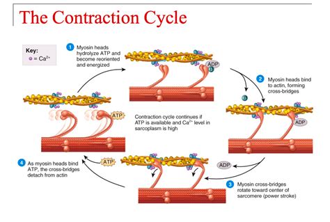
Table of Contents
The Functional Units of Contraction in a Muscle Fiber: Sarcomeres
Understanding how muscles contract requires delving into their fundamental building blocks. This article explores the sarcomere, the functional unit of contraction in a muscle fiber. We'll examine its structure, the proteins involved, the sliding filament theory, and the intricate processes that lead to muscle contraction and relaxation. By the end, you'll have a comprehensive understanding of this crucial biological mechanism.
The Sarcomere: A Microscopic Marvel
The sarcomere is the smallest functional unit of a myofibril, the rod-like structures that make up muscle fibers. These highly organized structures are responsible for the striated appearance of skeletal and cardiac muscle. Think of the sarcomere as the tiny engine driving muscle movement. Each sarcomere is defined by the Z-lines, or Z-discs, which act as anchors for the contractile proteins.
Components of the Sarcomere:
The sarcomere contains several key protein components, meticulously arranged to achieve efficient contraction:
-
Actin: A thin filamentous protein, arranged in a double helix structure. Actin filaments are anchored to the Z-lines. These filaments possess binding sites for myosin, the protein responsible for the power stroke during muscle contraction.
-
Myosin: A thick filamentous protein, with a head and tail region. The myosin heads possess ATPase activity, enabling them to hydrolyze ATP, providing the energy for muscle contraction. The myosin heads project outward from the thick filaments, forming cross-bridges with the actin filaments.
-
Z-lines (Z-discs): These are dense protein structures that serve as the boundaries of the sarcomere. Actin filaments are attached to the Z-lines.
-
M-line: Located in the center of the sarcomere, the M-line is a protein structure that anchors the myosin filaments. It ensures proper alignment of the thick filaments during contraction.
-
I-band: The lighter band of the sarcomere, contains only thin filaments (actin). It lies between the A-band and the Z-line. The I-band shortens during muscle contraction.
-
A-band: The darker band of the sarcomere, containing both thick (myosin) and thin (actin) filaments. The A-band remains relatively constant in length during muscle contraction.
-
H-zone: Located in the center of the A-band, the H-zone contains only thick filaments (myosin). This zone shortens during muscle contraction. It disappears completely during maximal contraction.
The Sliding Filament Theory: The Mechanism of Contraction
The sliding filament theory explains how muscle contraction occurs at the sarcomere level. This theory postulates that muscle contraction is achieved by the sliding of actin filaments over myosin filaments, resulting in the shortening of the sarcomere. This process is not about the filaments themselves shortening, but about their relative positions changing.
Steps in Muscle Contraction:
-
Excitation-Contraction Coupling: A nerve impulse triggers the release of acetylcholine (ACh) at the neuromuscular junction. ACh binds to receptors on the muscle fiber membrane, leading to depolarization and the release of calcium ions (Ca2+) from the sarcoplasmic reticulum (SR).
-
Calcium Binding: The released Ca2+ ions bind to troponin, a protein complex associated with actin. This binding causes a conformational change in troponin, which moves tropomyosin, another protein associated with actin.
-
Cross-Bridge Formation: The movement of tropomyosin exposes the myosin-binding sites on actin. Myosin heads then bind to these sites, forming cross-bridges.
-
Power Stroke: ATP hydrolysis by the myosin head provides the energy for the power stroke. The myosin head pivots, pulling the actin filament towards the center of the sarcomere.
-
Cross-Bridge Detachment: A new ATP molecule binds to the myosin head, causing it to detach from the actin filament.
-
ATP Hydrolysis and Re-attachment: The myosin head then hydrolyzes the ATP, returning to its high-energy conformation, ready to bind to another actin binding site further along the filament. This cycle repeats as long as Ca2+ and ATP are available.
-
Sarcomere Shortening: The repeated cycles of cross-bridge formation, power stroke, and detachment cause the actin filaments to slide over the myosin filaments, shortening the sarcomere and ultimately the entire muscle fiber.
-
Relaxation: When the nerve impulse ceases, Ca2+ is actively pumped back into the SR, reducing the cytosolic Ca2+ concentration. This leads to tropomyosin covering the myosin-binding sites on actin, preventing further cross-bridge formation and causing muscle relaxation.
Proteins Involved in Sarcomere Function: A Deeper Dive
Several other proteins play vital roles in the intricate orchestration of sarcomere function:
-
Titin: A giant protein that extends from the Z-line to the M-line, providing structural support to the sarcomere and acting as a molecular spring. It helps maintain the integrity of the sarcomere during contraction and relaxation.
-
Nebulin: A protein that runs along the length of the actin filament, helping regulate its length and assembly.
-
Tropomyosin: A protein that wraps around the actin filament, covering the myosin-binding sites in the absence of calcium.
-
Troponin: A protein complex consisting of three subunits (troponin I, troponin T, and troponin C). Troponin C binds to Ca2+, initiating the conformational changes that expose the myosin-binding sites on actin.
Types of Muscle Contractions: Isometric and Isotonic
Muscle contractions can be categorized into two main types:
-
Isometric Contractions: These involve muscle tension without a change in muscle length. Think of holding a heavy object in place – your muscles are working hard, generating force, but the length of the muscle doesn't change.
-
Isotonic Contractions: These involve muscle tension with a change in muscle length. There are two subtypes:
- Concentric Contractions: The muscle shortens while generating force (e.g., lifting a weight).
- Eccentric Contractions: The muscle lengthens while generating force (e.g., lowering a weight slowly).
Factors Affecting Muscle Contraction:
Several factors influence the strength and duration of muscle contractions:
-
Frequency of Stimulation: Increased frequency of nerve impulses leads to more frequent calcium release and more sustained contractions (tetanus).
-
Number of Motor Units Recruited: The more motor units recruited, the stronger the contraction.
-
Length of the Sarcomere: Optimal sarcomere length allows for maximal overlap between actin and myosin filaments, resulting in the strongest contraction. Too much or too little overlap reduces the efficiency of contraction.
-
ATP Availability: ATP is essential for both muscle contraction (power stroke) and relaxation (calcium pump). Depletion of ATP leads to muscle fatigue.
Muscle Fatigue: A Breakdown in the System
Muscle fatigue occurs when the muscle's ability to generate force decreases. Several factors contribute to muscle fatigue, including:
-
Depletion of Energy Stores: Reduced ATP and glycogen levels.
-
Accumulation of Metabolic Byproducts: Build-up of lactic acid and other metabolic wastes.
-
Electrolyte Imbalances: Changes in the concentrations of ions like potassium and sodium.
-
Central Fatigue: A decrease in neural drive to the muscles.
Conclusion: The Sarcomere – A Masterpiece of Biological Engineering
The sarcomere, with its intricate arrangement of proteins and precise mechanisms, represents a remarkable example of biological engineering. Understanding its structure and function is fundamental to comprehending the complexities of muscle contraction, a process vital for movement, posture, and numerous other bodily functions. Further research continues to unravel the finer details of sarcomere function and its implications for health and disease. From the microscopic level of the sarcomere to the macroscopic actions of the whole muscle, the principles discussed here provide a solid foundation for understanding this essential biological process. The ongoing research in this field promises further insights into muscle physiology, potential therapies for muscle diseases, and a deeper appreciation of the intricate workings of the human body.
Latest Posts
Latest Posts
-
How Many Valence Electrons Are In P
Apr 08, 2025
-
What Is The Sum Of Potential And Kinetic Energy
Apr 08, 2025
-
What Are The Lengths Of Line Segments Ab And Bc
Apr 08, 2025
-
What Is The Name Of Mg No3 2
Apr 08, 2025
-
Atomic Number Is Equal To The Number Of Protons
Apr 08, 2025
Related Post
Thank you for visiting our website which covers about Name The Functional Units Of Contraction In A Muscle Fiber . We hope the information provided has been useful to you. Feel free to contact us if you have any questions or need further assistance. See you next time and don't miss to bookmark.