Labelled Diagram Of A Reflex Arc
News Leon
Mar 20, 2025 · 6 min read
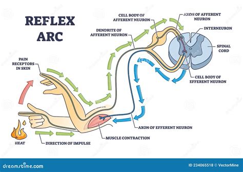
Table of Contents
The Reflex Arc: A Comprehensive Guide with Labelled Diagrams
The reflex arc is a neural pathway that controls a reflex action. It's a rapid, involuntary, and automatic response to a stimulus, bypassing the brain for incredibly fast reaction times. Understanding the reflex arc is crucial to understanding the basic workings of the nervous system. This comprehensive guide will explore the components, types, and significance of the reflex arc, accompanied by detailed labelled diagrams.
Components of the Reflex Arc
A typical reflex arc consists of five essential components:
-
Receptor: This is the specialized sensory nerve ending that detects the stimulus. Different receptors respond to different types of stimuli—pressure, temperature, light, or chemicals. For example, the receptors in your skin detect pain, heat, or pressure. These receptors are often located in the skin, muscles, tendons, or internal organs. They convert the stimulus into an electrical signal, initiating the reflex.
-
Sensory Neuron (Afferent Neuron): This neuron transmits the nerve impulse from the receptor to the central nervous system (CNS). The sensory neuron's axon carries the impulse towards the spinal cord or brainstem. The cell body of the sensory neuron is located outside the CNS, in a structure called the dorsal root ganglion.
-
Integration Center (Synapse): This is the point of connection between the sensory neuron and the motor neuron. It's located in the grey matter of the spinal cord or brainstem. Here, the signal is processed. In simple reflexes, this processing is minimal, involving only a single synapse. More complex reflexes may involve multiple synapses and interneurons.
-
Motor Neuron (Efferent Neuron): This neuron transmits the nerve impulse from the CNS to the effector. Its axon carries the impulse away from the CNS towards the muscle or gland. The cell body of the motor neuron is located in the anterior horn of the spinal cord's grey matter.
-
Effector: This is the muscle or gland that carries out the response to the stimulus. Muscles contract, causing movement, while glands secrete hormones or other substances. The effector's action is the observable reflex response.
Labelled Diagram of a Simple Reflex Arc (Monosynaptic Reflex)
Let's visualize the pathway with a diagram focusing on the knee-jerk reflex (patellar reflex), a classic example of a monosynaptic reflex arc (involving only one synapse):
                     +-----------------+
                     |     Receptor    |  (Muscle spindle in quadriceps)
                     +--------+--------+
                             |
                             | Sensory Neuron (Afferent)
                             v
             +-----------------+-----------------+
             |                 |                 |
             | Dorsal Root     | Spinal Cord     |
             | Ganglion       | Grey Matter     |
             +--------+--------+                 |
                             |                 |  Synapse
                             |                 |
                             | Motor Neuron (Efferent)
                             v
             +-----------------+-----------------+
             |                 |                 |
             |  Ventral Root  |                 |
             +--------+--------+                 |
                             |
                             |
                     +-----------------+
                     |     Effector     |  (Quadriceps muscle)
                     +-----------------+
Key:
- Receptor: Muscle spindle within the quadriceps muscle. This is sensitive to muscle stretch.
- Sensory Neuron: Carries the impulse from the muscle spindle to the spinal cord.
- Spinal Cord: The integration center where the sensory neuron synapses with the motor neuron.
- Motor Neuron: Carries the impulse from the spinal cord to the quadriceps muscle.
- Effector: Quadriceps muscle, which contracts in response to the stimulus.
Labelled Diagram of a Complex Reflex Arc (Polysynaptic Reflex)
Many reflexes involve more than one synapse, utilizing interneurons. Consider the withdrawal reflex (flexor reflex) as an example:
                         +-----------------+
                         |     Receptor    |  (Nociceptor in skin)
                         +--------+--------+
                                 |
                                 | Sensory Neuron (Afferent)
                                 v
                +-----------------+-----------------+
                |                 |                 |
                | Dorsal Root     | Spinal Cord     |
                | Ganglion       | Grey Matter     |
                +--------+--------+                 |
                                 |                 | Synapse 1
                                 |                 | (Sensory to Interneuron)
                                 | Interneuron       |
                                 |                 | Synapse 2
                                 |                 | (Interneuron to Motor Neuron)
                                 v                 |
                +-----------------+-----------------+
                |                 |                 |
                |  Ventral Root  |                 |
                +--------+--------+                 |
                                 |
                                 | Motor Neuron (Efferent) to Flexor Muscle
                                 v
                +-----------------+-----------------+
                |                 |                 |
                |    Flexor       |                 |  (Biceps) Contracts
                |     Muscle      |                 |
                +-----------------+-----------------+

                         +-----------------+
                         |    Motor Neuron | (to Extensor)
                         |    (Efferent)   |
                         +--------+--------+
                                 |
                +-----------------+-----------------+
                |                 |                 |
                |    Extensor      |                 |  (Triceps) Relaxes
                |     Muscle      |
                +-----------------+-----------------+
Key:
- Receptor: Nociceptor (pain receptor) in the skin.
- Sensory Neuron: Carries the impulse from the nociceptor to the spinal cord.
- Interneuron: Relays the signal to both flexor and extensor motor neurons.
- Motor Neuron (Flexor): Carries the impulse to the flexor muscle (e.g., biceps).
- Motor Neuron (Extensor): Carries the impulse to the extensor muscle (e.g., triceps).
- Effectors: Flexor and extensor muscles; the flexor contracts, and the extensor relaxes, causing withdrawal of the limb.
Types of Reflex Arcs
Reflex arcs are categorized based on the number of synapses involved and the location of integration:
-
Monosynaptic Reflexes: Involve only one synapse between the sensory and motor neuron. The knee-jerk reflex is a classic example. These are the fastest reflexes.
-
Polysynaptic Reflexes: Involve two or more synapses, with one or more interneurons. The withdrawal reflex is a prime example. These reflexes are slower than monosynaptic reflexes due to the additional synaptic delays.
-
Cranial Reflexes: The integration center is in the brainstem. Examples include pupillary reflexes and gag reflex.
-
Spinal Reflexes: The integration center is in the spinal cord. The knee-jerk and withdrawal reflexes are spinal reflexes.
Significance of the Reflex Arc
The reflex arc is vital for several reasons:
-
Protection: It provides rapid protection from harmful stimuli, like withdrawing from a hot stove or blinking to protect the eyes.
-
Homeostasis: Reflexes help maintain internal balance. Examples include reflexes controlling heart rate, blood pressure, and breathing.
-
Posture and Balance: Reflexes involving muscles and joints help maintain posture and balance.
-
Coordination of Movement: Reflexes are fundamental for coordinated movement, enabling smooth, precise actions.
-
Diagnosis: Testing reflexes is crucial in neurological examinations. Abnormal reflexes can indicate damage to the nervous system. The absence or exaggeration of a reflex can pinpoint the location of neurological injury or disease.
Clinical Significance of Reflex Arc Assessment
Assessing reflex responses forms a cornerstone of neurological examination. Clinicians use a reflex hammer to elicit reflexes and observe the response's speed, strength, and symmetry. Abnormalities might indicate:
-
Upper Motor Neuron Lesions: Causes hyperreflexia (exaggerated reflexes) and clonus (rhythmic muscle contractions).
-
Lower Motor Neuron Lesions: Causes hyporeflexia (diminished or absent reflexes) and muscle atrophy.
-
Peripheral Neuropathy: Affects the sensory or motor neurons, leading to altered reflexes.
-
Spinal Cord Injuries: Can cause either hyperreflexia or hyporeflexia below the level of the injury.
-
Metabolic Disorders: Some metabolic disorders can affect nerve function, resulting in altered reflexes.
Conclusion
The reflex arc is a fundamental neural pathway essential for survival and maintaining bodily function. Understanding its components, types, and clinical significance is crucial for appreciating the complexity and elegance of the nervous system. The labelled diagrams provide a visual representation of these intricate processes, allowing for a deeper understanding of the fast and efficient responses that protect us and maintain homeostasis. Furthermore, the clinical implications of reflex arc assessment highlight the importance of these involuntary actions in diagnosing various neurological conditions. Continued study and exploration of the reflex arc offer valuable insights into the intricate workings of the human body.
Latest Posts
Latest Posts
-
Which Three Dimensional Figure Is Formed By The Rotation Given
Mar 21, 2025
-
75 Percent Of What Number Is 15
Mar 21, 2025
-
Time Magazine Person Of The Century 1999
Mar 21, 2025
-
Ground State Electron Configuration For Titanium
Mar 21, 2025
-
Which Of The Following Is The Most Acidic
Mar 21, 2025
Related Post
Thank you for visiting our website which covers about Labelled Diagram Of A Reflex Arc . We hope the information provided has been useful to you. Feel free to contact us if you have any questions or need further assistance. See you next time and don't miss to bookmark.
