Label The Structures Of A Nephron In The Figure
News Leon
Mar 19, 2025 · 5 min read
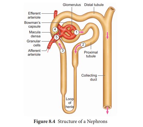
Table of Contents
Label the Structures of a Nephron: A Comprehensive Guide
The nephron, the functional unit of the kidney, is a complex structure responsible for filtering blood and producing urine. Understanding its intricate anatomy is crucial for grasping the physiological processes involved in maintaining fluid and electrolyte balance, blood pressure regulation, and waste excretion. This article provides a detailed guide to labeling the structures of a nephron, accompanied by explanations of their functions and significance. We'll explore the nephron's components in detail, covering both cortical and juxtamedullary nephrons, and highlighting their key differences.
The Major Components of a Nephron: A Visual Journey
Before we dive into the specifics, let's establish a foundational understanding. A nephron consists of two main parts: the renal corpuscle and the renal tubule. Imagine the renal corpuscle as the initial filtration unit, while the renal tubule fine-tunes the filtrate, reabsorbing essential substances and secreting waste products.
1. The Renal Corpuscle: The Filtration Starting Point
The renal corpuscle, located in the cortex of the kidney, is responsible for the initial filtration of blood. It comprises two structures:
-
Glomerulus: A network of capillaries where blood filtration occurs. The glomerular capillaries are fenestrated, meaning they have pores allowing for the passage of water and small solutes while retaining larger molecules like proteins and blood cells. Think of it as a highly selective sieve. High glomerular capillary pressure is crucial for effective filtration.
-
Bowman's Capsule (Glomerular Capsule): A double-walled epithelial cup surrounding the glomerulus. The inner layer of Bowman's capsule, consisting of specialized cells called podocytes, plays a significant role in filtration. Podocytes have intricate foot processes that interdigitate, forming filtration slits that further refine the filtrate. The outer layer of Bowman's capsule is a parietal layer. The space between the two layers is called the Bowman's space, where the filtered fluid (glomerular filtrate) collects.
2. The Renal Tubule: Refining the Filtrate
The renal tubule is a long, convoluted tube where the initial filtrate undergoes further modification before becoming urine. It consists of several distinct segments:
-
Proximal Convoluted Tubule (PCT): This segment is characterized by its extensive length and brush border, which greatly increases its surface area for absorption and secretion. The PCT is the primary site for the reabsorption of essential nutrients, such as glucose, amino acids, and water. It also actively secretes substances like hydrogen ions (H+) and certain drugs. Remember the high capacity for reabsorption within the PCT.
-
Loop of Henle (Nephron Loop): This U-shaped structure extends from the cortex into the medulla. It plays a crucial role in concentrating urine. The descending limb is permeable to water but relatively impermeable to solutes, while the ascending limb is impermeable to water but actively transports solutes out of the tubule. The countercurrent multiplier system, involving the loop of Henle and the vasa recta (peritubular capillaries), is essential for establishing the concentration gradient in the medulla. The length of the loop of Henle varies significantly between cortical and juxtamedullary nephrons, contributing to the kidney's ability to produce concentrated urine.
-
Distal Convoluted Tubule (DCT): This segment is shorter than the PCT and is less involved in reabsorption. However, it plays a vital role in regulating electrolyte balance, particularly calcium, sodium, and potassium. The DCT is also sensitive to hormonal regulation, responding to hormones like aldosterone and parathyroid hormone. Focus on the hormonal regulation of the DCT.
-
Connecting Tubule and Collecting Duct: The connecting tubule connects the DCT to the collecting duct. Multiple nephrons share a single collecting duct. The collecting duct system plays a significant role in regulating water balance and urine concentration under the influence of antidiuretic hormone (ADH). The regulation of water permeability by ADH is paramount here.
Cortical vs. Juxtamedullary Nephrons: A Comparative Analysis
While all nephrons share the basic structural components, there are two main types based on their location and the length of their Loop of Henle:
Cortical Nephrons: These nephrons are predominantly located in the cortex of the kidney, with short loops of Henle that barely penetrate the medulla. They primarily focus on filtration and reabsorption of essential nutrients and electrolytes.
Juxtamedullary Nephrons: These nephrons have long loops of Henle that extend deep into the medulla. Their longer loops of Henle are crucial for establishing the concentration gradient necessary for producing concentrated urine, particularly important during dehydration.
The Juxtaglomerular Apparatus (JGA): A Specialized Region
The JGA is a specialized structure located at the junction of the afferent arteriole, efferent arteriole, and distal convoluted tubule. It plays a critical role in regulating blood pressure and glomerular filtration rate (GFR). The JGA comprises several cell types:
-
Juxtaglomerular Cells (JG cells): Modified smooth muscle cells in the afferent arteriole that secrete renin, a crucial enzyme in the renin-angiotensin-aldosterone system (RAAS), which regulates blood pressure.
-
Macula Densa Cells: Specialized epithelial cells in the distal convoluted tubule that detect changes in sodium concentration and fluid flow in the tubule. They signal the JG cells to adjust renin secretion.
-
Extraglomerular Mesangial Cells: These cells are located between the afferent and efferent arterioles and are thought to have a role in communication between the macula densa and JG cells.
Clinical Significance: Understanding Nephron Dysfunction
Understanding the structure and function of the nephron is crucial in diagnosing and managing various kidney diseases. Nephron damage can lead to a range of conditions, including:
-
Acute Kidney Injury (AKI): Sudden loss of kidney function, often due to factors such as dehydration, infections, or medications.
-
Chronic Kidney Disease (CKD): Progressive loss of kidney function over time, often due to conditions like diabetes and hypertension.
-
Glomerulonephritis: Inflammation of the glomeruli, often caused by autoimmune diseases or infections.
-
Polycystic Kidney Disease (PKD): Genetic disorder characterized by the development of cysts in the kidneys, leading to impaired kidney function.
Conclusion: Mastering the Nephron's Anatomy
This comprehensive guide offers a detailed walkthrough of the nephron's structures, emphasizing their functions and clinical relevance. By understanding the intricate interplay between the renal corpuscle and the renal tubule, and appreciating the differences between cortical and juxtamedullary nephrons, you gain a deeper insight into the complexities of renal physiology. This knowledge forms a strong foundation for comprehending kidney function in health and disease. Remember to consult reputable anatomical resources for visual aids and further detailed information. Mastering the nephron’s structure is key to understanding the crucial role of the kidneys in maintaining overall health.
Latest Posts
Related Post
Thank you for visiting our website which covers about Label The Structures Of A Nephron In The Figure . We hope the information provided has been useful to you. Feel free to contact us if you have any questions or need further assistance. See you next time and don't miss to bookmark.