Av Valves Prevent Backflow Into The
News Leon
Mar 28, 2025 · 6 min read
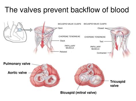
Table of Contents
AV Valves: Preventing Backflow and Ensuring Efficient Blood Circulation
The heart, a tireless powerhouse, relentlessly pumps blood throughout our bodies. This intricate process relies heavily on a sophisticated system of valves, ensuring unidirectional blood flow. Among these crucial components are the atrioventricular (AV) valves, critical players in preventing backflow and maintaining the efficiency of the circulatory system. This comprehensive article will delve into the structure, function, and significance of AV valves, exploring their role in preventing backflow and the consequences of their malfunction.
Understanding the Atrioventricular Valves: Structure and Location
The human heart possesses four valves, strategically positioned to regulate blood movement between its chambers and the major blood vessels. The two AV valves, the mitral (bicuspid) and tricuspid valves, are located between the atria and ventricles, acting as one-way gates.
The Mitral Valve: Guardian of the Left Atrium-Ventricular Junction
Situated between the left atrium and the left ventricle, the mitral valve is a bicuspid valve, meaning it comprises two cusps or leaflets. These leaflets, composed of tough, fibrous connective tissue covered by endocardium (the innermost layer of the heart), open to allow blood to flow from the left atrium into the left ventricle during diastole (the relaxation phase of the heart). During systole (the contraction phase), the mitral valve closes tightly, preventing the backflow of blood from the left ventricle into the left atrium. This precise action is crucial for maintaining efficient blood flow to the systemic circulation (the body's circulatory system).
The Tricuspid Valve: Regulating Flow into the Right Ventricle
Positioned between the right atrium and the right ventricle, the tricuspid valve, as its name suggests, possesses three cusps. Similar in structure to the mitral valve, its leaflets are composed of connective tissue and endocardium. The tricuspid valve's function mirrors that of the mitral valve: it opens to allow blood flow from the right atrium to the right ventricle during diastole and closes tightly during systole to prevent backflow into the right atrium. This prevents recirculation and ensures that blood flows efficiently towards the pulmonary circulation (the lungs).
The Mechanics of AV Valve Closure: Preventing Backflow
The precise opening and closing of the AV valves are orchestrated by a complex interplay of pressure gradients and specialized structures:
Pressure Gradients: The Driving Force
The primary mechanism controlling AV valve function is the pressure difference between the atria and ventricles. During diastole, when the atria are filled with blood, atrial pressure exceeds ventricular pressure, causing the AV valves to open passively. Blood flows freely from the atria to the ventricles. Conversely, during systole, ventricular pressure rises dramatically as the ventricles contract. This increase in ventricular pressure surpasses atrial pressure, forcing the AV valves to close. The leaflets come together, effectively sealing the opening between the atria and ventricles.
Papillary Muscles and Chordae Tendineae: The Anchoring System
To ensure complete closure and prevent the leaflets from inverting (prolapsing) into the atria under the high pressure of ventricular contraction, the AV valves are anchored by a system of papillary muscles and chordae tendineae. Papillary muscles are small muscles protruding from the ventricular walls. Chordae tendineae, strong fibrous cords, connect the papillary muscles to the edges of the AV valve leaflets. When the ventricles contract, the papillary muscles contract simultaneously, tightening the chordae tendineae and preventing leaflet prolapse. This intricate system is vital for maintaining the structural integrity of the AV valves and preventing backflow.
Consequences of AV Valve Dysfunction: Backflow and Heart Failure
When AV valves malfunction, they fail to effectively prevent backflow, leading to a range of cardiovascular complications. The most common causes of AV valve dysfunction include:
Mitral Valve Prolapse (MVP): A Common Cause of Backflow
MVP refers to the displacement of one or both mitral valve leaflets into the left atrium during ventricular contraction. This prolapse can lead to mitral regurgitation (leakage of blood back into the left atrium), reducing the efficiency of blood flow to the systemic circulation. MVP can be asymptomatic or cause symptoms such as palpitations, shortness of breath, and chest pain.
Mitral Stenosis: Narrowed Opening, Reduced Blood Flow
Mitral stenosis is a condition characterized by the narrowing of the mitral valve opening. This narrowing restricts blood flow from the left atrium to the left ventricle, increasing pressure in the left atrium and potentially leading to pulmonary congestion (fluid buildup in the lungs). Symptoms can include fatigue, shortness of breath, and palpitations.
Tricuspid Regurgitation: Leakage in the Right Side of the Heart
Similar to mitral regurgitation, tricuspid regurgitation involves the leakage of blood back into the right atrium from the right ventricle during ventricular contraction. This can lead to reduced efficiency in the pulmonary circulation and symptoms such as fatigue, edema (swelling), and palpitations.
Tricuspid Stenosis: Obstruction of Right-Sided Flow
Tricuspid stenosis, less common than mitral stenosis, involves the narrowing of the tricuspid valve orifice. This obstruction reduces blood flow from the right atrium to the right ventricle, potentially leading to right-sided heart failure. Symptoms can include peripheral edema, jugular venous distension, and ascites (abdominal fluid accumulation).
Diagnosing AV Valve Dysfunction: Identifying the Problem
Diagnosing AV valve dysfunction typically involves a combination of techniques:
Physical Examination: Listening for Murmurs
A physical examination, including auscultation (listening to the heart sounds with a stethoscope), can reveal abnormal heart sounds, such as murmurs, indicative of valve dysfunction. These murmurs result from turbulent blood flow due to backflow or stenosis.
Echocardiography: Visualizing Valve Structure and Function
Echocardiography, an ultrasound technique, provides detailed images of the heart's structure and function. It allows clinicians to visualize the AV valves, assess their opening and closing, and detect any abnormalities, such as prolapse, stenosis, or regurgitation.
Cardiac Catheterization: Invasive Assessment
In some cases, cardiac catheterization, a more invasive procedure, may be necessary to obtain precise measurements of pressure gradients across the AV valves and assess the severity of dysfunction.
Treating AV Valve Dysfunction: Restoring Normal Blood Flow
Treatment options for AV valve dysfunction vary depending on the severity of the condition and the presence of symptoms.
Medical Management: Addressing Symptoms
For mild cases of AV valve dysfunction, medical management may focus on symptom relief using medications such as diuretics (to reduce fluid retention), anticoagulants (to prevent blood clots), and heart failure medications.
Surgical Intervention: Valve Repair or Replacement
Severe AV valve dysfunction often necessitates surgical intervention. Valve repair aims to restore normal valve function without replacing the valve. Valve replacement involves replacing the damaged valve with a prosthetic valve, either mechanical or biological.
Conclusion: The Vital Role of AV Valves in Cardiovascular Health
The atrioventricular valves are essential components of the circulatory system, playing a pivotal role in preventing backflow and ensuring efficient blood circulation. Their precise opening and closing mechanisms are finely tuned to maintain unidirectional blood flow between the atria and ventricles. Dysfunction of these valves can have serious consequences, potentially leading to heart failure and other cardiovascular complications. Early diagnosis and appropriate management are crucial to prevent or mitigate the impact of AV valve disorders, maintaining cardiovascular health and overall well-being. Further research continues to improve our understanding of AV valve dysfunction and to develop innovative treatment strategies. This ongoing pursuit of knowledge aims to enhance the quality of life for individuals affected by these critical cardiovascular conditions. Understanding the complexities of AV valve function underscores the remarkable sophistication of the human cardiovascular system and highlights the importance of maintaining heart health.
Latest Posts
Latest Posts
-
The Height Above Sea Level Is Called
Mar 31, 2025
-
For What Value Of X Is Abc Def
Mar 31, 2025
-
Which Elements Have 5 Valence Electrons
Mar 31, 2025
-
Which Of The Following Is An Example Of Nonvolatile Memory
Mar 31, 2025
-
How To Calculate Voltage Of A Cell
Mar 31, 2025
Related Post
Thank you for visiting our website which covers about Av Valves Prevent Backflow Into The . We hope the information provided has been useful to you. Feel free to contact us if you have any questions or need further assistance. See you next time and don't miss to bookmark.
