Atrioventricular Valves Prevent Backflow Into The
News Leon
Mar 26, 2025 · 6 min read
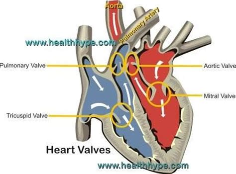
Table of Contents
Atrioventricular Valves: Preventing Backflow and Ensuring Efficient Cardiac Function
The human heart, a tireless engine driving life's processes, relies on a complex interplay of chambers, vessels, and valves to ensure unidirectional blood flow. Central to this intricate system are the atrioventricular (AV) valves, crucial structures preventing the backflow of blood and maintaining the efficiency of the cardiac cycle. Understanding their structure, function, and the consequences of their malfunction is vital for appreciating the intricacies of cardiovascular health.
The Anatomy of Atrioventricular Valves: A Closer Look
The heart possesses two AV valves: the tricuspid valve, located between the right atrium and right ventricle, and the mitral valve (bicuspid valve), situated between the left atrium and left ventricle. These valves, unlike the semilunar valves (pulmonary and aortic), are characterized by their unique anatomical features:
Tricuspid Valve:
- Three cusps (leaflets): Unlike the mitral valve, the tricuspid valve comprises three cusps of fibrous tissue, each anchored by chordae tendineae.
- Chordae tendineae: These strong, fibrous cords attach the cusps to the papillary muscles, preventing valve prolapse during ventricular contraction.
- Papillary muscles: These muscles, projecting from the ventricular wall, contract synchronously with the ventricles, tightening the chordae tendineae and maintaining valve closure.
- Location: Strategically placed between the right atrium and right ventricle, it regulates blood flow from the atrium to the ventricle.
Mitral Valve:
- Two cusps (leaflets): The mitral valve, also known as the bicuspid valve, consists of two leaflets, anterior and posterior.
- Chordae tendineae: Similar to the tricuspid valve, strong chordae tendineae connect the cusps to the papillary muscles.
- Papillary muscles: These muscles, located within the left ventricle, coordinate their contraction with the ventricle's contraction to prevent mitral valve prolapse.
- Location: Positioned between the left atrium and left ventricle, ensuring unidirectional blood flow from the atrium to the ventricle.
The importance of these structural components cannot be overstated. The coordinated action of the cusps, chordae tendineae, and papillary muscles ensures that the valves open effectively to allow blood flow during diastole (relaxation) and close tightly during systole (contraction) to prevent backflow. This precision is essential for maintaining efficient cardiac output.
The Physiology of Atrioventricular Valve Function: A Symphony of Movement
The function of the AV valves is intimately linked to the cardiac cycle, the rhythmic sequence of contraction and relaxation that propels blood throughout the body. The valves' actions can be broken down into two phases:
Valve Opening (Diastole):
During diastole, the ventricles relax, and the pressure within them falls below the atrial pressure. This pressure difference causes the AV valves to open passively. Blood flows freely from the atria into the ventricles, filling them in preparation for the next contraction. The cusps are open, allowing unimpeded blood flow.
Valve Closing (Systole):
As the ventricles contract during systole, the intraventricular pressure rises dramatically. This increased pressure pushes the AV valve cusps together, causing them to close. Simultaneously, the papillary muscles contract, tightening the chordae tendineae. This prevents the cusps from inverting (prolapsing) into the atria, maintaining the valve's integrity and preventing backflow. The precise timing and coordination of these events are critical for efficient cardiac function.
Consequences of Atrioventricular Valve Dysfunction: A Cascade of Problems
Malfunction of the AV valves, whether due to congenital defects, rheumatic heart disease, or degenerative changes, can lead to a range of serious cardiovascular complications:
Mitral Valve Prolapse (MVP):
MVP occurs when one or both mitral valve leaflets bulge back into the left atrium during ventricular contraction. This can cause a heart murmur and, in severe cases, lead to heart failure. While many individuals with MVP are asymptomatic, others may experience palpitations, shortness of breath, or chest pain.
Mitral Valve Regurgitation (MVR):
MVR, also known as mitral insufficiency, occurs when the mitral valve doesn't close properly, allowing blood to leak back from the left ventricle into the left atrium during systole. This reduces the amount of blood effectively pumped to the body, leading to symptoms such as fatigue, shortness of breath, and eventually heart failure.
Tricuspid Valve Regurgitation (TVR):
Similar to MVR, TVR involves the incomplete closure of the tricuspid valve, causing blood to leak back from the right ventricle into the right atrium during systole. This condition can lead to symptoms such as fatigue, edema (swelling), and shortness of breath.
Mitral Stenosis:
Mitral stenosis is a narrowing of the mitral valve opening, hindering blood flow from the left atrium to the left ventricle. This increases the pressure in the left atrium, leading to symptoms like shortness of breath, fatigue, and palpitations. Severe stenosis can result in pulmonary edema and heart failure.
Tricuspid Stenosis:
Tricuspid stenosis is a less common condition involving the narrowing of the tricuspid valve opening. This obstructs blood flow from the right atrium to the right ventricle, causing similar symptoms to mitral stenosis, but often affecting the systemic circulation less dramatically in the early stages.
Diagnosis and Treatment of Atrioventricular Valve Disorders: Modern Approaches
Diagnosing AV valve disorders typically involves a combination of physical examination, electrocardiogram (ECG), echocardiogram, and cardiac catheterization. The choice of treatment depends on the severity of the condition and the individual's overall health:
Medical Management:
For mild cases, medical management may involve medications to control symptoms and reduce the workload on the heart. Diuretics can help reduce fluid buildup, while other medications can manage irregular heart rhythms.
Surgical Intervention:
In more severe cases, surgical intervention may be necessary. Options include:
- Valve repair: This involves surgically repairing the damaged valve to restore its function. Valve repair is generally preferred over replacement whenever feasible, preserving the patient's native valve and avoiding the need for lifelong anticoagulation.
- Valve replacement: This involves replacing the damaged valve with a prosthetic valve, either mechanical or biological. Mechanical valves are durable but require lifelong anticoagulation therapy to prevent blood clots. Biological valves are less durable but do not require lifelong anticoagulation.
The advancements in cardiac surgery have significantly improved the outcomes of AV valve replacement and repair, providing patients with a better quality of life and increased longevity.
Conclusion: The Vital Role of Atrioventricular Valves in Cardiovascular Health
The atrioventricular valves play a pivotal role in the efficient functioning of the heart. Their precise opening and closing ensure unidirectional blood flow, preventing backflow and maintaining optimal cardiac output. Understanding their structure, function, and the consequences of their dysfunction is crucial for appreciating the complexity and importance of cardiovascular health. Advances in diagnostic techniques and surgical interventions offer hope for individuals affected by AV valve disorders, improving their prognosis and enhancing their quality of life. Continued research into the prevention and treatment of these conditions remains essential for safeguarding cardiovascular health. Staying informed and proactive about your heart health is paramount in maintaining well-being and preventing future complications. Regular checkups, healthy lifestyle choices, and prompt medical attention when necessary can significantly reduce the risk of developing AV valve disorders and improve the overall cardiovascular health.
Latest Posts
Latest Posts
-
Regulates What Enters And Leaves The Cell
Mar 29, 2025
-
The Most Active Phagocytic Cells In Circulating Blood Are
Mar 29, 2025
-
How Many Cups In 9 Ounces
Mar 29, 2025
-
How Many Ones Are There Between 1 And 100
Mar 29, 2025
-
025 Expressed As A Percentage Is
Mar 29, 2025
Related Post
Thank you for visiting our website which covers about Atrioventricular Valves Prevent Backflow Into The . We hope the information provided has been useful to you. Feel free to contact us if you have any questions or need further assistance. See you next time and don't miss to bookmark.
