Animal Vs Plant Cell Venn Diagram
News Leon
Mar 16, 2025 · 6 min read
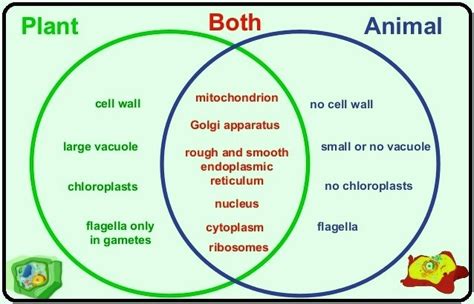
Table of Contents
Animal vs. Plant Cell Venn Diagram: A Comparative Analysis of Eukaryotic Cells
Understanding the fundamental differences and similarities between animal and plant cells is crucial for grasping the basics of biology. While both are eukaryotic cells—meaning they possess a membrane-bound nucleus and other organelles—they exhibit distinct characteristics reflecting their different functions and lifestyles. A Venn diagram provides a visually compelling way to compare and contrast these cellular structures. This article delves deep into the specifics of animal and plant cells, using a Venn diagram as a framework for understanding their unique features and shared components.
The Core Components: What Animal and Plant Cells Share
Both animal and plant cells are eukaryotic, meaning they possess a complex internal organization with membrane-bound organelles. This shared characteristic sets them apart from prokaryotic cells like bacteria, which lack such compartmentalization. The Venn diagram's overlapping section represents these common features:
1. Cell Membrane (Plasma Membrane):
The cell membrane, a selectively permeable barrier, regulates the passage of substances into and out of the cell. It’s composed primarily of a phospholipid bilayer, embedded with proteins and cholesterol. This dynamic structure maintains cellular integrity and facilitates communication with the external environment. Both animal and plant cells rely on their cell membrane for nutrient uptake, waste removal, and signal transduction.
2. Cytoplasm:
The cytoplasm is the gel-like substance filling the cell, excluding the nucleus. It's a dynamic environment where many metabolic processes occur. Ribosomes, the protein synthesis machinery, are suspended within the cytoplasm in both cell types. The cytoskeleton, a network of protein filaments, provides structural support and facilitates intracellular transport in both animal and plant cells. This shared cytoplasm provides a vital medium for cellular activities.
3. Nucleus:
The nucleus is the control center of the cell, housing the genetic material (DNA) organized into chromosomes. It’s enclosed by a double membrane called the nuclear envelope, punctuated by nuclear pores that regulate the transport of molecules between the nucleus and cytoplasm. Both cell types utilize the nucleus for DNA replication, transcription (creating RNA from DNA), and regulating gene expression, essential for cell function and survival.
4. Mitochondria:
The powerhouse of the cell, mitochondria are responsible for cellular respiration—the process of generating ATP (adenosine triphosphate), the cell's primary energy currency. They are double-membrane-bound organelles containing their own DNA and ribosomes, a vestige of their endosymbiotic origin. Both animal and plant cells rely heavily on mitochondria to fuel their various metabolic processes.
5. Endoplasmic Reticulum (ER):
The ER is a network of interconnected membranes extending throughout the cytoplasm. It exists in two forms: rough ER (studded with ribosomes) involved in protein synthesis and modification, and smooth ER, which plays a role in lipid synthesis, detoxification, and calcium storage. Both animal and plant cells utilize the ER for these crucial biosynthetic and metabolic functions.
6. Golgi Apparatus (Golgi Body):
The Golgi apparatus is a stack of flattened membrane sacs that processes, modifies, sorts, and packages proteins and lipids received from the ER. It plays a key role in directing these molecules to their final destinations within or outside the cell. Both animal and plant cells utilize the Golgi to ensure proper protein trafficking and secretion.
7. Ribosomes:
As previously mentioned, ribosomes are the protein synthesis factories of the cell. These complex molecular machines translate the genetic code from mRNA (messenger RNA) into polypeptide chains, which fold into functional proteins. Both animal and plant cells require ribosomes for protein production, essential for all cellular processes.
8. Lysosomes (Animal Cells) / Vacuoles (Plant Cells):
While functionally analogous, the structures differ slightly. Animal cells possess lysosomes, membrane-bound organelles containing hydrolytic enzymes that break down cellular waste and debris. Plant cells, on the other hand, primarily utilize vacuoles for storage, waste breakdown, and maintaining turgor pressure. Both structures contribute to cellular maintenance and waste management.
9. Peroxisomes:
Peroxisomes are small, membrane-bound organelles that participate in various metabolic reactions, including the breakdown of fatty acids and detoxification of harmful substances. Both animal and plant cells utilize peroxisomes for these important metabolic roles.
Unique Features: Diverging Paths of Animal and Plant Cells
The sections of the Venn diagram not overlapping represent the features unique to each cell type. These differences reflect their distinct roles in multicellular organisms.
Plant Cell-Specific Features:
1. Cell Wall: A rigid outer layer surrounding the plant cell membrane, the cell wall provides structural support, protection, and maintains cell shape. It’s composed primarily of cellulose, a complex carbohydrate. The cell wall is crucial for plant cells to withstand turgor pressure and maintain their upright form.
2. Chloroplasts: These are the sites of photosynthesis, the process of converting light energy into chemical energy in the form of glucose. Chloroplasts contain chlorophyll, the green pigment that captures light energy, and are crucial for plant growth and survival as they are the primary producers in most ecosystems. They are double-membrane-bound organelles with their own DNA and ribosomes, like mitochondria.
3. Large Central Vacuole: Plant cells possess a large, central vacuole that occupies a significant portion of the cell's volume. This vacuole plays a crucial role in maintaining turgor pressure, storing water, nutrients, and waste products, and regulating cellular pH.
Animal Cell-Specific Features:
1. Centrioles: These cylindrical structures, made of microtubules, play a crucial role in cell division, specifically organizing the mitotic spindle during mitosis and meiosis. While some plant cells might possess centrioles, they are generally absent in higher plant cells and are not essential for cell division in most plant species.
2. Flagella/Cilia: These are motile appendages that enable some animal cells to move. Flagella are long, whip-like structures, while cilia are shorter and hair-like. They are involved in locomotion and also in moving substances across the cell surface. While some plant cells possess flagella in their reproductive cells (e.g., sperm), they are much less common in plants than in animals.
3. Lysosomes (primary): While plant cells have vacuoles that perform similar functions, animal cells use lysosomes more extensively for intracellular digestion and waste breakdown.
The Venn Diagram in Action: A Visual Representation
The information above can be beautifully organized into a Venn diagram. Imagine two overlapping circles. One circle represents "Animal Cell," and the other represents "Plant Cell."
-
Overlapping Section (Both Animal and Plant Cells): Cell membrane, cytoplasm, nucleus, mitochondria, endoplasmic reticulum, Golgi apparatus, ribosomes, peroxisomes.
-
Animal Cell-Specific Section: Centrioles, flagella/cilia, lysosomes (primary).
-
Plant Cell-Specific Section: Cell wall, chloroplasts, large central vacuole.
Conclusion: Understanding the Nuances of Cellular Diversity
The Venn diagram provides a powerful visual tool for comparing and contrasting animal and plant cells. While they share fundamental eukaryotic characteristics, their unique features reflect their specialized roles and adaptations within their respective environments. Understanding these similarities and differences is fundamental to appreciating the diversity of life and the intricate workings of cellular biology. This knowledge forms a crucial base for further studies in cell biology, genetics, and other related biological fields. The differences highlighted here are not absolute; exceptions exist within the vast diversity of plant and animal life. However, the general principles illustrated by the Venn diagram provide a valuable framework for understanding cellular organization and function. Further research into specific plant and animal cell types can reveal even more nuanced differences and similarities.
Latest Posts
Latest Posts
-
Is A Webcam An Input Or Output Device
Mar 17, 2025
-
Word For A Person Who Uses Big Words
Mar 17, 2025
-
What Is 375 As A Percentage
Mar 17, 2025
-
Which Is The Correct Order Of The Scientific Method
Mar 17, 2025
-
How Long Is A Thousand Days
Mar 17, 2025
Related Post
Thank you for visiting our website which covers about Animal Vs Plant Cell Venn Diagram . We hope the information provided has been useful to you. Feel free to contact us if you have any questions or need further assistance. See you next time and don't miss to bookmark.
