Agarose Gel Electrophoresis Separates Nucleic Acid Fragments According To
News Leon
Apr 03, 2025 · 7 min read

Table of Contents
Agarose Gel Electrophoresis: Separating Nucleic Acid Fragments by Size and Charge
Agarose gel electrophoresis is a fundamental technique in molecular biology used to separate nucleic acid fragments—DNA or RNA—based on their size and charge. This powerful method allows researchers to analyze, visualize, and quantify nucleic acids, making it indispensable in various applications, from genetic fingerprinting to gene cloning and disease diagnostics. Understanding the principles behind agarose gel electrophoresis is crucial for interpreting results and effectively employing this technique in research settings.
The Principles of Separation: Size and Charge
The separation of nucleic acid fragments in agarose gel electrophoresis relies on two primary factors: size and charge. Nucleic acids, due to their phosphate backbone, carry a uniform negative charge at neutral or alkaline pH. When placed in an electric field, these negatively charged molecules migrate towards the positive electrode (anode). However, their movement isn't uniform.
The Sieving Effect of Agarose
The agarose gel itself acts as a molecular sieve. The gel matrix consists of a porous network of agarose fibers. Smaller fragments can navigate this network more easily than larger fragments. Larger fragments experience more resistance and move slower through the gel matrix. This size-dependent migration is the primary factor responsible for the separation of fragments.
Influence of Voltage and Runtime
The applied voltage also influences the separation. A higher voltage accelerates the migration of all fragments, but it can also lead to heating and potential distortion of the bands. Therefore, an optimal voltage is chosen to balance speed and resolution. The electrophoresis runtime is adjusted to allow sufficient separation of the fragments based on their size.
Components of Agarose Gel Electrophoresis
Several essential components are involved in the successful execution of agarose gel electrophoresis:
1. Agarose Gel
Agarose is a polysaccharide extracted from seaweed. It forms a gel matrix when dissolved in a suitable buffer and allowed to solidify. The concentration of agarose determines the pore size of the gel. Higher agarose concentrations (e.g., 1-2%) produce gels with smaller pores, suitable for separating smaller DNA fragments. Lower concentrations (e.g., 0.5-0.8%) create larger pores, ideal for resolving larger fragments. The choice of agarose concentration is critical for optimizing the separation.
2. Electrophoresis Buffer
The buffer provides ions to carry the current and maintains the pH of the gel during electrophoresis. Common buffers include Tris-acetate-EDTA (TAE) and Tris-borate-EDTA (TBE). TBE buffer has a higher buffering capacity, which is advantageous for longer runs, but TAE buffer is preferred for some applications because it gives less background signal in downstream processes such as DNA purification.
3. DNA Sample
The DNA sample must be prepared appropriately before loading onto the gel. This typically involves mixing the DNA with a loading dye, which contains:
-
Tracking dye: A colored dye that allows you to monitor the progress of the electrophoresis. Bromophenol blue and xylene cyanol are common tracking dyes. These dyes migrate at specific rates through the gel.
-
Density agent: Glycerol or sucrose increase the density of the sample, allowing it to sink into the wells of the gel.
4. Electrophoresis Chamber
The electrophoresis chamber houses the gel and provides electrodes for applying the electric field. It's filled with electrophoresis buffer to ensure proper electrical conductivity.
5. Power Supply
The power supply provides the electric current to drive the electrophoresis. The voltage and runtime are controlled using the power supply.
Steps in Performing Agarose Gel Electrophoresis
-
Gel Preparation: Weigh the required amount of agarose and dissolve it in the appropriate volume of electrophoresis buffer by heating (usually in a microwave). Once dissolved, the solution is poured into a casting tray with a comb to create wells. Allow the gel to solidify.
-
Gel Loading: Carefully remove the comb and submerge the gel in the electrophoresis chamber filled with electrophoresis buffer. Load the DNA samples into the wells using a micropipette. Load a DNA ladder (a mixture of DNA fragments of known sizes) into one of the wells for size comparison.
-
Electrophoresis: Connect the electrophoresis chamber to the power supply and apply the appropriate voltage. The electrophoresis runs until the tracking dye has migrated far enough down the gel.
-
Visualization: After electrophoresis, the DNA fragments are invisible to the naked eye. To visualize them, the gel is stained with a DNA-binding dye, usually ethidium bromide or a safer alternative like SYBR Safe. Ethidium bromide intercalates between DNA base pairs and fluoresces under UV light. This allows the visualization of DNA bands.
-
Analysis: The separated DNA fragments appear as distinct bands on the gel. The size of each fragment can be estimated by comparing its migration distance to that of the DNA ladder. The intensity of each band reflects the amount of DNA in that fragment. Gel documentation systems capture images of the stained gel for analysis and record-keeping.
Applications of Agarose Gel Electrophoresis
Agarose gel electrophoresis has numerous applications in various fields of molecular biology and related disciplines. Some notable applications include:
1. DNA Fingerprinting and Forensic Science
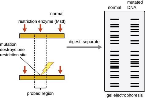
Agarose gel electrophoresis is used to separate DNA fragments generated by restriction enzyme digestion. The resulting pattern of fragments is unique to an individual and serves as a DNA fingerprint, valuable in forensic investigations for identifying suspects or victims.
2. Gene Cloning and Recombinant DNA Technology
Verification of successful gene cloning often involves analyzing the size of the cloned DNA fragment through agarose gel electrophoresis. This helps confirm that the gene of interest has been inserted into the vector.
3. PCR Product Analysis
Polymerase chain reaction (PCR) is a technique widely used to amplify specific DNA sequences. Agarose gel electrophoresis is routinely used to analyze the size and quantity of PCR products.
4. RNA Analysis
Agarose gel electrophoresis can also be used to separate RNA fragments. This is useful for analyzing RNA integrity and for studying gene expression levels. The use of denaturing agarose gels (containing formaldehyde or glyoxal) is often necessary to separate RNA fragments based on size alone, as RNA molecules can adopt secondary structures.
5. Disease Diagnostics
Agarose gel electrophoresis is used in various diagnostic applications, including the detection of genetic mutations associated with certain diseases. For example, it can be used to detect the presence of specific DNA sequences indicative of certain pathogens.
6. Quality Control
Agarose gel electrophoresis is important in various laboratory settings for quality control. This includes checking the integrity of DNA or RNA samples before downstream processes, such as sequencing, or confirming the success of a cloning experiment.
Advantages and Limitations of Agarose Gel Electrophoresis
Advantages:
- Relatively simple and inexpensive: The equipment and materials required are readily available and relatively inexpensive.
- Versatile: It can be used to separate a wide range of nucleic acid fragment sizes.
- High resolution: Agarose gels can provide good resolution of DNA fragments, enabling accurate size determination.
- Easy visualization: DNA fragments can be easily visualized using DNA-binding dyes.
Limitations:
- Limited resolution for very small or very large fragments: Agarose gels are not suitable for resolving very small (<100 bp) or very large (>20 kb) DNA fragments. Different techniques, such as capillary electrophoresis for small fragments and pulsed-field gel electrophoresis for large fragments, are used instead.
- Ethidium bromide is a mutagen: While safer alternatives exist, traditional DNA stains like ethidium bromide are potential mutagens, requiring careful handling and disposal.
- Time-consuming: The process can be time-consuming, especially for longer electrophoresis runs.
Conclusion
Agarose gel electrophoresis remains a cornerstone technique in molecular biology laboratories worldwide. Its simplicity, cost-effectiveness, and versatility make it an indispensable tool for analyzing and manipulating nucleic acids. While limitations exist, the technique’s widespread applications across diverse research areas reaffirm its enduring importance in advancing our understanding of genetic information and its application in different fields, from fundamental research to clinical diagnostics. Understanding the principles of the technique and its proper execution is crucial for obtaining reliable and interpretable results.
Latest Posts
Latest Posts
-
Which Type Of Connective Tissue Is Avascular
Apr 04, 2025
-
Ionic Equation For Naoh And Hcl
Apr 04, 2025
-
Examples Of Gas In Everyday Life
Apr 04, 2025
-
Is The Human Body A Conductor
Apr 04, 2025
-
80 Of 45 Is What Number
Apr 04, 2025
Related Post
Thank you for visiting our website which covers about Agarose Gel Electrophoresis Separates Nucleic Acid Fragments According To . We hope the information provided has been useful to you. Feel free to contact us if you have any questions or need further assistance. See you next time and don't miss to bookmark.
